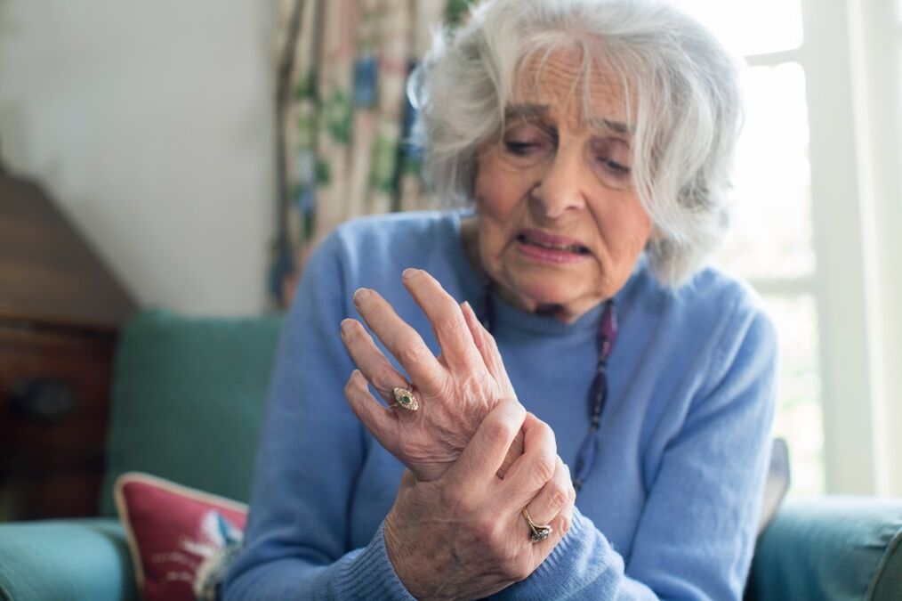
Joint arthrosis (also in the literature you can find the name "deforming arthrosis", "osteoarthritis" or "osteoarthritis") is a chronic degenerative process of joint tissue, in which, due to dystrophic processes, over time, cartilage covering the articular surface is destroyed. , in addition, the degenerative process can cover the actual joint capsule and bone tissue, causing bone deformation.
Types of joint arthrosis
In general, the term "arthrosis" is used to refer to a fairly large group of joint diseases, the mechanism of development of such pathology may differ to some extent. Often, you can experience arthrosis of large joints, this group of diseases includes:
- gonarthrosis - deformed lesions of the knee joint;
- coxarthrosis - pathology of the hip joint;
- arthrosis of the shoulder joint;
- arthrosis of the elbow joint, etc.
Less frequently arthrosis of small joints develops: hands (more often interphalangeal and metacarpophalangeal) and feet. In addition, spondyloarthrosis is differentiated - dystrophic lesions of the intervertebral joints, referred to as spinal diseases, although the mechanism of its development is similar to other types of arthrosis. If the pathology spreads to several joints, we can say about the general arthrosis, or - polyarthrosis.
In medicine, it is customary to distinguish two main types of joint arthrosis, depending on the mechanism of its development. Primary arthrosis (also called idiopathic) is a pathology that develops mainly in the joint tissues, beyond any deviations in the systemic work of the body. Primary arthrosis is characterized by an increase in degenerative processes in cartilage tissue simultaneously with a violation of its recovery.
Secondary arthrosis is the result of injury to a joint due to trauma (traumatic arthrosis). In addition, certain pathological processes in the body, in particular, disorders of mineral metabolism and others, can lead to the development of secondary arthrosis.
At a young age, as a rule, traumatic arthrosis is detected. In elderly patients, in some cases, it is impossible to distinguish between primary and secondary joint damage - closely related are many pathological processes in the body.

Why does arthrosis develop?
Although there is no agreement on the exact cause of the development of arthrosis, the factors that contribute to pathological changes in joint tissue are known to scientists. The main reason for the development of idiopathic joint arthrosis is a variety of hereditary and genetic factors. First of all, this group includes congenital features of the composition of joint tissues, which contribute to more intensive than normal cartilage destruction and its recovery is too slow. In addition, hereditary factors can include a variety of congenital malformations and deformities of the osteoarticular system (dysplasia of the joints, their excessive movement, deformities of the spine, legs, hands, etc. ), which are caused by excessive or non -physiological load placed on certain joints. , why the articular surface can develop improperly, deformed, and the cartilage that covers it - collapse;
The following reasons can lead to the development of secondary joint arthrosis:
- mechanical damage to the joint is the result of certain mechanical influences that cause a violation of the anatomical integrity of the structures that make up the joint. The mechanical injury group includes injuries, surgical interventions, excessive physical activity and sports;
- joint disease - first of all, pathology of inflammatory nature;
- metabolic disorders that lead to changes in the composition of the cartilage covering the articular surface, as it becomes more vulnerable, ruptures faster and recovers more slowly;
- some diseases of the endocrine glands, which can also lead to metabolic disorders;
- a number of autoimmune pathologies in which cells of the immune system invade the body’s own tissues, in this case, joint tissues, provoking destruction;
- vascular pathology, as a result of which the blood supply to the tissues is insufficient and the development of dystrophic processes in them.
How does joint arthrosis develop?
It is believed that the first process of destruction of the joint in joint arthrosis is the defeat of cartilage. It begins with a violation of microcirculation in the capillaries of the periosteum, which are located below the cartilage that covers the articular surface of the bone. Typically, nutrients in cartilage tissue come from articular fluid and from nearby bone tissue. When blood circulation is disrupted in the periosteum ducts, cartilage nutrition is disrupted. Gradually, cartilage tissue loses its natural elasticity, becomes thinner, its surface is uneven, microcracks and tubercles may appear on it, interfering with the sliding of the interconnected articular surfaces. The composition of synovial fluid will definitely change.
With movement in the joints, pain, itching, clicks begin to interfere. Over time, the pathology develops, the range of motion in the joint decreases, the joint gap becomes narrower, the cartilage on the protruding part of the articular surface can become thinner until it disappears completely, and osteophytes form at the edges of the articular surface.
The mechanism described is characteristic primarily of dementia arthrosis, which develops gradually over time. The mechanism of development of other forms of arthrosis - e. g. , post -traumatic, postoperative, post -infectious, associated with metabolic disorders - may be slightly different, but in general, changes in joints with such pathology are similar to dementia arthrosis.
Stages of arthrosis and manifestations
The specific manifestation of the pathological process, the severity of which depends greatly on how strong the process of destruction in the joint, how much tissue is involved in it. Even so, there are two main clinical symptoms of arthrosis at any stage. First of all, it is joint pain. In addition, decreased joint mobility is a concern.
In our country, it is customary to distinguish the degree of arthrosis in accordance with the clinical and radiological classifications applied in 1961.
Arthrosis of the joints 1 degree- the early stages of development of pathological processes.The main symptom is stiff joint movements in the morning, after resting. As soon as the patient begins to move, the stiffness disappears. In the joints, there may be a slight disturbance of movement, minor pain may be interrupted after being at rest, during the first movement. Fractures in the joints are often observed. However, there is no obvious pain after performing normal movements, the pain can appear only with a clear load on the joints, and after rest disappears by itself.
X-ray examination of the affected joint did not show significant changes in anatomical structure; in some cases, there may be a slight narrowing of the joint space or the presence of small -sized single bone growths along the edges of the articular surface.
Because there is no obvious pain and movement in the joints, patients rarely seek medical help at this stage of disease development.
2 degrees of arthrosis - the development of the disease.This is indicated by the appearance of severe and acute pain, as well as different irritations, by pressing on the joint during movement in it. The range of movement in the joints is very limited, therefore, if we talk about the defeat of large joints, the possibility of shortening of limb function may occur. The pain bothers in the morning, immediately after waking up, as in the first stage of arthrosis, but in contrast, they are stronger, longer, often they gradually turn into daytime pain. The latter is formed during the day, when mechanical work on the joints gradually decreases the ability of cartilage amortization. At this stage, there is a fairly significant destruction of the joint, a deformation of the bone that forms it. Joint meteosensitivity may interfere: the appearance or significant increase in pain as the weather changes, which is associated with a decrease in the compensatory properties of joint tissue and its ability to regulate intra-articular pressure during atmospheric pressure fluctuations.
X-ray examination showed a significant narrowing of the joint gap, a large amount of bone growth. Bone tissue in the pericartylaginous zone is closed due to obvious dystrophic processes; cyst cavities can form in them.
At this stage of the disease, the patient’s ability to work is reduced; he was unable to do any number of jobs at all because of severe pain during movement on the affected joint or its contractures.
Grade 3 joint arthrosis corresponds to an advanced stage of the pathological process.Pain is always disturbing - both during movement and in a state of complete rest, which is associated with a number of factors: inflammation of the joint tissue, deformation of the articular surface, spasm of the surrounding muscles. Its range of motion is very limited, in some cases, generally impossible. Movements in the affected joints are accompanied by strong cramps, not only audible to the patient, but also to those around him. The joint at this stage is significantly deformed, fluid accumulates in the joint capsule due to an intense inflammatory process. Severe meteosensitivity develops: intensification of pain due to climate change. Muscles in large joint areas are spasmodic, due to lack of mobility, their atrophy can develop. With degree 3 arthrosis of the knee joint, curvature of the foot can be observed - varus (in the form of the letter "O") or valgus (in the form of the letter "X").
X-ray images showed an almost complete absence of joint space, the articular surface was significantly deformed, and large bone growths were located at its edges. Intra-articular structures (menisci, ligaments) are destroyed, bone tissue is slipped. Tissue around the joint is calcified, articular rats can appear in the joint cavity - fragments of bone tissue.
With third -degree joint arthrosis, there is a constant decrease in the patient’s ability to work, his disability.
4 degrees of arthrosis - the degree of complete destruction of the affected joint.His "restriction" grew - the impossibility of even the slightest movement due to severe pain. The pain is insurmountable even with strong non -narcotic analgesics, they cannot be eliminated with physiotherapy procedures. With the defeat of the knee joint, the patient loses the ability to move freely. Strong inflammation of the joint tissues can trigger joint consolidation (ankylosis) or the formation of false joints (neoarthrosis).
On the roentgenogram, one can see the strongest sclerosis of the bone forming the joint, its fusion, a large number of osteophytes, and strong calcification of the joint tissue.
How is arthrosis treated?
The scope of therapeutic measures for the treatment of arthrosis of the joints depends on the stage of the disease and the prevalence of pathological processes. There is a simple pattern: the earlier arthrosis treatment is started, the more effective it is. Therefore, it is very important to see a doctor on time, at the first signs of discomfort in the joint area, at the moment of morning cramps or the appearance of cramps during movement. In the early stages of the pathological process, effectively take vitamin-mineral complexes and chondroprotectors-drugs that increase metabolism in cartilage tissue and its structure. For example, a drug whose active ingredient is crystalline glucosamine sulfate - a natural component of healthy cartilage. It stimulates the production of proteoglycans and at the same time inhibits the process of destruction of cartilage tissue.

A good addition to the listed measures to treat arthrosis is physiotherapy exercises, physiotherapy techniques, it is important to follow a diet with adequate amounts of protein and fat in the diet. Timely therapeutic and prophylactic measures make it possible not only to eliminate pain and discomfort, but also to prevent the development of the disease, the pathological transition to a more severe level.
Second degree and more severe arthrosis cannot be completely cured. At this stage, the treatment of arthrosis is reduced to the elimination or reduction of pain, as well as the emphasis of inflammation on the joint tissue. To relieve pain, non-steroidal anti-inflammatory drugs and non-narcotic analgesics are used, in the form of local agents (ointments, gels with anesthetic and anti-inflammatory effects), as well as systemic drugs. An important role is played by relieving the load on the diseased joint, which makes it possible to reduce mechanical damage to the articular surface.
Once the acute pain is eliminated, an important task is to normalize metabolic processes in cartilage tissue, slowing down the destructive processes in it, which is also recommended to take chondroprotective drugs. Also, drugs that stimulate tissue microcirculation have proven themselves well. At the stage of remission of arthrosis (if there is no severe pain), physiotherapy training, physiotherapy is useful, which will reduce the likelihood of other exacerbations and reduce the need for analgesics and NSAIDs.
The list of physiotherapeutic measures in the treatment of joint arthrosis includes electrophoresis, ultrasound exposure, radiation and magnetic therapy. The most important condition for the effectiveness of such measures is to carry out physiotherapeutic treatment of arthrosis only in the period outside the exacerbation of the disease, otherwise there is a high probability of activating the inflammatory process and increasing pain.
How to treat arthrosis in the fourth stage of the disease? Joint tissue at this stage is practically destroyed, the only effective solution is surgical intervention, in which the damaged joint is replaced with a special endoprosthesis. Not necessarily, such an operation allows you to fully restore movement in the joints, however, after the completion of the recovery period, the patient can return to active, work, relieve pain.
Arthrosis, whose symptoms and treatment are described in this article, is a serious pathology that can lead to disability, significantly reducing its quality of life. Timely treatment initiated can slow the progression of the disease, prevent the development of severe complications, and maintain the patient’s activity and ability to work.


